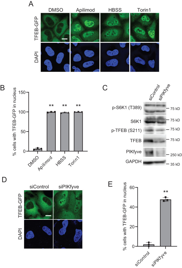
FIGURE 2:
PIKfyve inhibition induces the dephosphorylation of TFEB and nuclear translocation of TFEB. (A) HeLa cells stably expressing TFEB-GFP treated with the indicated drugs were cultured for 1 h in the growth medium or HBSS for 1 h. Cells were fixed and stained with DAPI for nuclear staining and then analyzed by immunofluorescence microscopy. Bar, 10 µm. (B) Percentage of TFEB-GFP that is localized to the nucleus in A (mean ± SD; n > 100 cells from three independent experiments). **P < 0.01. (C) HeLa cells were transfected with siControl or siPIKfyve for 72 h and then analyzed by immunoblot using the indicated antibodies. (D) HeLa cells stably expressing TFEB-GFP were transfected with siControl or siPIKfyve for 72 h. Cells were fixed and stained with DAPI for nuclear staining and then analyzed by immunofluorescence microscopy. Bar, 10 µm. (E) Percentage of TFEB-GFP that is localized to the nucleus in D (mean ± SD; n > 50 cells from three independent experiments). **P < 0.01.

