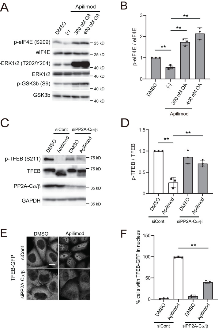
FIGURE 4:
PP2A knockdown suppresses PIKfyve-inhibition-dependent dephosphorylation and nuclear translocation of TFEB. (A) HeLa cells treated with 1 µM apilimod (or DMSO vehicle control) and okadaic acid at the indicated concentration were cultured for 1 h in the growth medium and then analyzed by immunoblot using the indicated antibodies. (B) Quantitation of phospho-elF4E signal intensities from immunoblots in A, following normalization to the total elF4E protein (mean ± SD; three independent experiments). **P < 0.01. (C) HeLa cells transfected with siControl, siPP2A-Cα and siPP2A-Cβ were treated with 1 µM apilimod (DMSO as a control) for 2 h and then analyzed by immunoblot using the indicated antibodies. (D) Quantitation of phospho-TFEB signal intensities from immunoblots in C, following normalization to the total TFEB protein (mean ± SD; three independent experiments). **P < 0.01. (E) HeLa cells transfected with siControl, siPP2A-Cα, and siPP2A-Cβ were treated with 1 µM apilimod (or DMSO vehicle control) for 2 h. Cells were fixed and then analyzed by immunofluorescence microscopy. Bar, 10 µm. (F) Percentage of TFEB-GFP that is localized to the nucleus in E (mean ± SD; n > 50 cells from three independent experiments). **P < 0.01.

