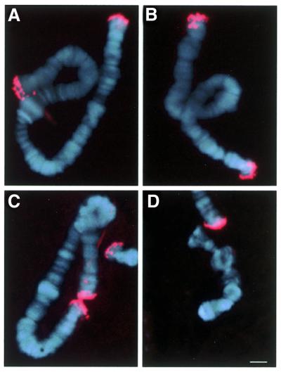
Figure 3.
In situ hybridisation experiments with a 29 bp oligonucleotide containing the HSE (see Materials and Methods) detected with an anti-digoxigenin–TRIC conjugate antibody. A compiled image with Hoechst H-33528 staining shows the signal obtained for (A) chromosome I, (B) chromosome II, (C) chromosome III and (D) chromosome IV. The bar represents 10 µm.

