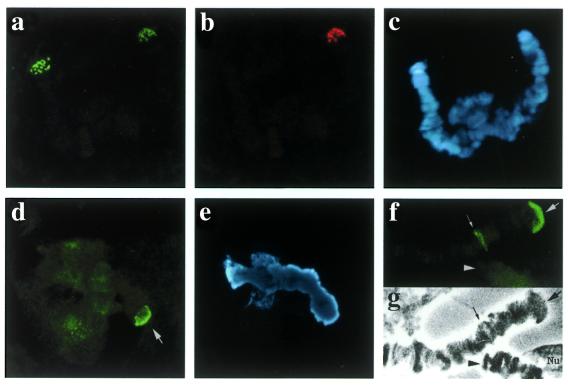
Figure 5.
Double in situ hybridisation in chromosome IV from C.piger × C.thummi hybrids with the telomeric 176 repeat labelled with 11-dUTP-biotin and detected with FITC (green) (a) and with a telomeric HSE oligonucleotide labelled with 11-dUTP-DIG and detected with TRIC (red) (b). In (c) Hoechst H-33258 staining shows the two chromatids unpaired, as frequently occurs. Immunohistochemical visualisation of HSF in heat shocked (1 h, 35°C) cells from C.piger: (d) Chromosome IV telomere 4R (arrow); (e) the corresponding Hoechst H-33258 image. HSF immunolocalization in heat shocked (2 h, 35°C) C.thummi larva: (f) positive signal at TBR III (3R telomere) (arrow) and heat shock puff III-A3 (small arrow) (note the absence of signal at the 4R end; arrowhead); (g) the corresponding phase contrast image.

