. 2022 Apr 15;22(3):e28. doi: 10.4110/in.2022.22.e28

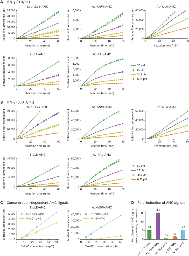
Figure 3. Measurement of i-proteasome activity in WCLs using fluorogenic reporter substrates. (A) The hydrolysis of fluorogenic peptide substrates (final 6.25, 12.5, 25, and 50 µM in 100 µl hydrolysis reaction) was continuously measured every 3 min for 60 min. WCLs (10 µl, corresponding up to 15 µg total protein) from untreated HeLa cells were added at time 0. Representative raw fluorescence graphs from three independent experiments are shown. (B) The hydrolysis of peptide substrates described in (A) was measured in WCLs from IFN-γ-treated HeLa cells (500 U/ml for 48 h). (C) The fluorescence intensities of the c-proteasome substrate (Z-LLE-AMC) and i-proteasome substrate (Ac-ANW-AMC) at 25 min time point; data are presented as the mean±SD (n=3). (D) The i-proteasome specificity of the X-AMC substrates was evaluated at 12.5 µM concentration in IFN-γ-treated samples. The fold induction of AMC fluorescence intensity over IFN-γ-untreated controls is shown.

Figure 3