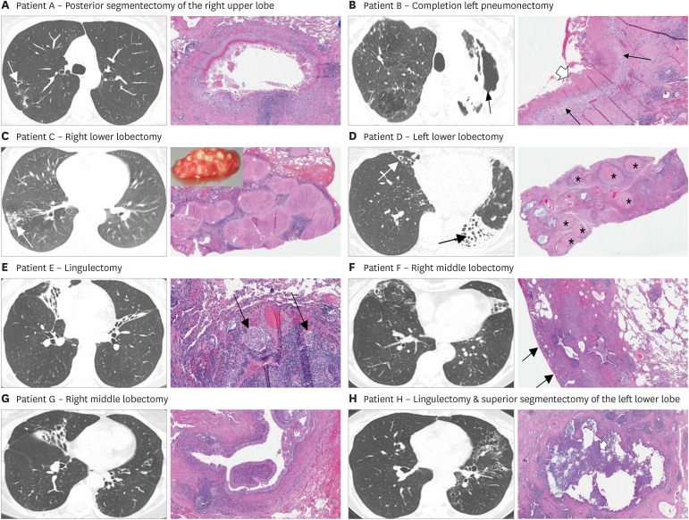
Figure 1. Representative axial CT image and the key histopathologic photomicrograph (H&E) for six patients with NTM lung disease. (A) Axial CT image shows a small cavity in the posterior segment of the right upper lobe and some surrounding tree-in-bud opacities. The lung photomicrograph demonstrates necrotizing granuloma with central necrotic material surrounded by epithelioid histiocytes, multinucleated giant cells and chronic inflammation, including lymphoid aggregates. (B) Axial CT image shows severe left upper lobe fibrocavitary disease with significant volume loss. The lung photomicrograph demonstrates necrotizing granuloma with layers of necrosis (thick white arrow), epithelioid histiocytes (thin black arrows) and surrounding fibrosis and chronic inflammation. (C) Axial CT image shows a bronchiectatic airway in the right lower lobe with numerous coalescing small to medium size nodules distally (arrow). The lung photomicrograph demonstrates numerous coalescing, necrotizing granulomas. Inset is the gross surgical tissue sample analyzed demonstrating the visible nodular lesions that corresponded to both the nodules found on the CT scan and the relatively large, nodular granulomas seen on histopathology. (D) Axial CT image shows severe bronchiectasis and atelectasis of the left lower lobe (black arrow) and moderate bronchiectasis in the right middle lobe (white arrow). The lung photomicrograph (low power) demonstrates numerous necrotizing granuloma (asterisks) with central necrosis surrounded by fibrosis and chronic inflammation. (E) Axial CT image shows severe bronchiectasis and atelectasis in the right middle lobe and in the superior segment of the lingula. The lung photomicrograph demonstrates an airway wall with chronic inflammation that includes lymphoid aggregates and nonnecrotizing granulomas with multinucleated giant cells (black arrows). (F) Axial CT image shows bronchiectasis and tree-in-bud opacities of the right middle lobe and atelectasis/bronchiectasis of the lingula. The lung photomicrograph demonstrates peripheral area of fibrosis (note the pleural surface—thin black arrows) surrounding mildly dilated small airway with lymphoid aggregates in the airway wall. Representative axial CT image and the key histopathologic photomicrograph (H&E) for two patients with chronic PsA-LD. (G) Axial CT image shows bronchiectasis of the central airways and the right middle lobe. The lung photomicrograph demonstrates dilated bronchiole, surrounded by collagenous fibrosis. (H) Axial CT image shows diffuse bronchiectasis of the left upper lobe and central region of the left lower lobe. The lung photomicrograph demonstrates a dilated small airway with inflammatory debris within the lumen (white arrow) with peripheral area of dense fibrosis.
Additional CT images and lung photomicrographs are shown in Supplementary Figs. 1, 2, 3, 4, 5, 6, 7, 8.

