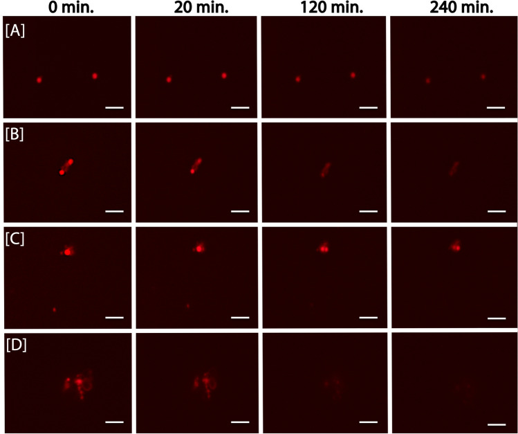
Fig. 4.
Snapshots of time-lapse microscopy of ANME enrichment culture labeled by mcrA FISH-TAMB probes. BE326 BH2 ANME enrichments were incubated anaerobically with 1 µM FISH-TAMB probes targeting mcrA mRNA and subsequently imaged via spinning-disk fluorescence confocal microscopy. Micrographs were snapped every minute for 14 h with an exposure of 100 ms. Composite micrographs of brightfield and Cy5 channel represent the first 4-h observation of A single cells; B and C physically associated cells; and D cell aggregate. Scale bar 5 µm

