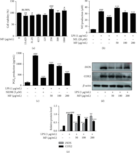
Figure 3.

Effect of MF on proinflammatory mediators in LPS-stimulated RAW264.7 macrophages. (a) The cells were treated with various concentrations of MF for 24 h, and their viability was determined by MTT assay. (b and c) The cells were treated with 50, 100, or 200 μg/mL of MF for 1 h prior to the addition of LPS (1 μg/mL) and were incubated for 24 h. NO and PGE2 levels were determined using Griess reagent and EIA kit, respectively. NIL (20 μM) and NS398 (5 μM) were used as positive controls for NO and PGE2 inhibition, respectively. (d and e) The protein levels of iNOS and COX2 were determined by western blot with specific antibodies. Densitometric analysis was performed using ImageJ software. The data are presented as the mean ± SD. ##p < 0.01 and ###p < 0.001 vs. the control group; ∗p < 0.05, ∗∗p < 0.01, and ∗∗∗p < 0.001 vs. the LPS-treated group.
