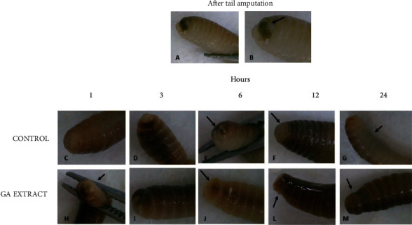
Figure 5.

Representative photographs of the surgical incision in the anterior segments of Eisenia fetida earthworms topically treated with GA-conjugated extract at 5 μg/mL concentration. Soon after the incision, the wound received a single topical dose of 2 μL of phosphate buffer (control) or extract solution. The earthworms were then kept in Petri dishes, and the regeneration process was evaluated in different periods of time. (a, b) Right after the incision, it is possible to observe the worm's intestinal tube (in yellowish-brown color indicated by the arrow). (c, h) One hour after cutting, the entire region was covered with mucus and seems to be more contracted in earthworms that received the extract treatment than in controls. (d, i) Three hours after cutting, the contraction of the wound is already well established and it is possible to see new transparent tissue starting to form. At some point, the wound is still open. Six hours after cutting, the wound still has open parts in the control worms (e, arrow) and is completely closed with new tissue in the worm topically treated with the extract (j, arrow); 12 hours after the incision, the wound of the control worms is completely regenerated with new whitish tissue (f, arrow). In earthworms treated with the extract, the regenerated area is already pigmenting and differentiating into the segments that have been cut (l, arrow). 24 h after the incision, the entire area was regenerated. However, pigmentation in the ventral region, which is slower than the dorsal region, has not yet occurred in the control earthworms (g, arrow), while in those treated topically with the extract, pigmentation is practically complete both in the dorsal region and in the ventral region of the animal's body (m, arrow).
