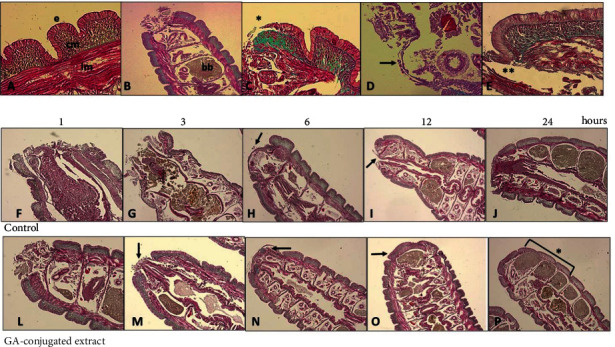
Figure 6.

Representative histological analysis of Eisenia fetida earthworms submitted to posterior surgical incision of the three last segments with and without topical treatment using GA-conjugated extract at 5 μg/mL concentration. The slides were stained with Masson-Goldner trichrome kit that is used for visualization of muscles, collagen fibers, connective tissues, and keratin. (a) Body wall histology showing the (e) epidermic layer that is directly connected with circular muscle layer (cm) immersed in an extracellular matrix richest in collagen. Immediately afterwards, there is the longitudinal muscular layer (lm) that is covered with the peritoneum that delimits the earthworm's coelomic cavity. (b) Posterior segments (metameres) showing the region of the surgical incision. In this place, there are spaces full of brown bodies (bb), which are structures that contain immune cells (cellomyocytes) that trapped and destroyed pathogens or linked to impurities and underwent melanization. When these cavities are completely filled, the worm performs autotomy to free them and then regenerates the posterior region. (c, d) Detail of the incision site showing migration (∗) of circular muscle cells migrating to initially close the wound. These cells behave similarly to human fibroblasts when skin damage occurs. (e) Only after migration and onset of proliferation of the smooth muscle layer does epidermis proliferation occur. (f–j) Sequence of healing processes in control earthworms at 1.3, 6, 12, and 24 hours. After this period, the region is in the final stage of regeneration of the incised segments. (l–p) Sequence of healing processes in earthworms topically treated with GA-conjugated extract showing acceleration in regenerative processes in relation to control. After 24 hours, the segments are fully regenerated (p, ∗).
