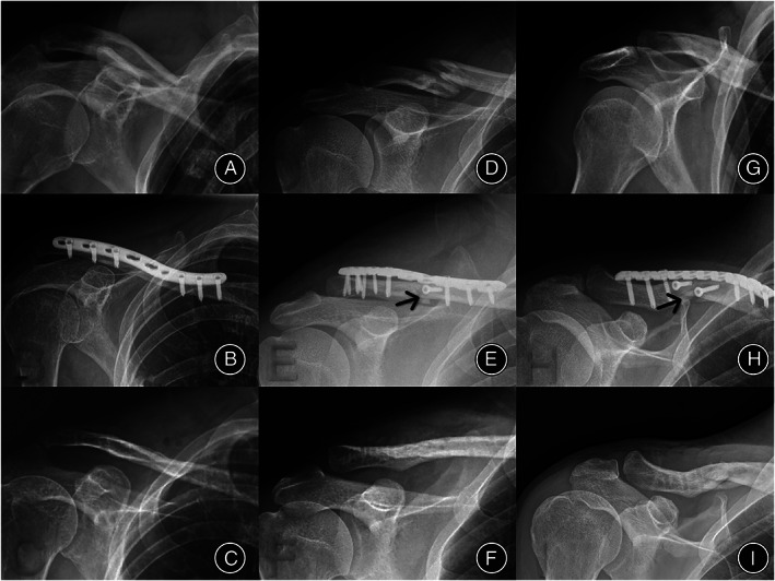
Fig. 3.

Radiographic findings of the healing process of three cases with different internal fixation methods. (A–C) Images of the SPF group before operation, after operation, and during fracture healing after removal of the internal fixation device, respectively. (D–F) Images of the PLFP group before operation, after operation, and during fracture healing after removal of the internal fixation device, respectively. The black mark in panel E indicates that the fracture end was vertically fixed outside the plate with a screw. (G–I) Images of the PLFP group before operation, after operation, and during fracture healing after removal of the internal fixation device, respectively. The black mark in panel H indicates that the fracture end is vertically fixed outside the plate with two screws. (C, F, and I) The fracture was healed well after the removal of internal fixation devices in both groups. Abbreviations: PLFP, plate combined with fracture local fixation; SPF, simple plate fixation
