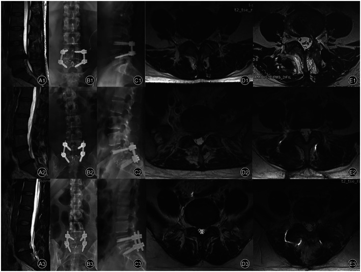
Fig. 7.

(A1–E1) One case of Wiltse approach TLIF patient. (A2–E2) One case of MIS‐TLIF patient, (A3–E3) One case of PLIF patient. (A1) MRI before surgery showed disc herniation in L4‐L5 segment. (B1 and C1) Postoperative anterior and lateral DR. (D1) Axial T2 MRI show disc herniation and multifidus before surgery. (E1) Axial T2 MRI show surgery level multifidus at last follow‐up after surgery with no significant atrophy and fatty infiltration. (A2) MRI before surgery showed disc herniation in L5‐S1 segment. (B2 and C2) postoperative anterior and lateral DR. (D2) Axial T2 MRI show disc herniation and multifidus before surgery. (E2) Axial T2 MRI show surgery level multifidus at last follow‐up after surgery with no significant atrophy and fatty infiltration. (A3) MRI before surgery showed disc herniation in L5‐S1 segment. (B3 and C3) Postoperative anterior and lateral DR. (D3) Axial T2 MRI show disc herniation and multifidus before surgery. (E3) Axial T2 MRI show surgery level multifidus at last follow‐up after surgery with significant atrophy and fatty infiltration.
