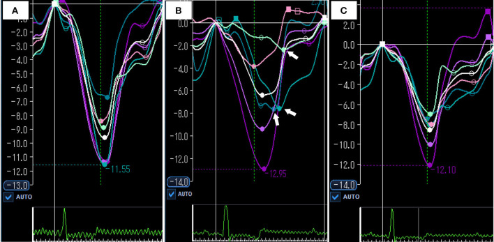
Figure 4.
Representative images of speckle tracking echocardiography at baseline (A) acute period (B) and compensation period (C). (A) Global RVLS was −9.6% and RV-SD6 was 16.8 msec at baseline. (B) In acute period, global RVLS was significantly impaired compared with baseline (– 6.4%). Systolic shortening times of free wall were delayed (white arrows), and RV-SD6 was increased (81.5 msec). (C) In compensation period, global RVLS and RV-SD6 were returned to the baseline level (−8.6% and 10.1 msec). White color curve indicates global RVLS. RVLS, right ventricular longitudinal strain; RV-SD6, standard deviation of systolic shortening time of right ventricular six segments.

