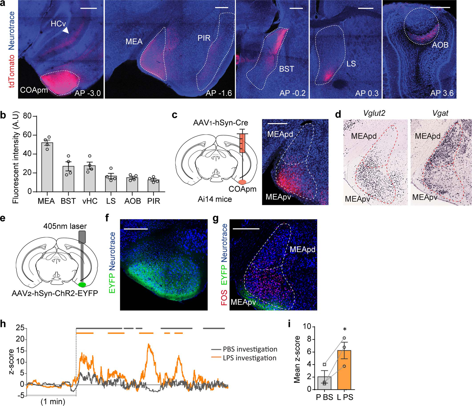
Extended Data Figure 6. COApm neurons preferentially project to MEA-Vglut2(+) neurons.

a-b, Virus encoding the anterograde tracer (AAV2-hSyn-tdTomato) was targeted to the COApm. Total fluorescence intensity was measured in sub-regions receiving COApm axonal projections: MEA, BST, ventral hippocampus (HCv), lateral septum (LS), AOB and piriform cortex (PIR) (n=4 mice; from 2 independent experiments). Scale bar=500μm. c, Virus encoding the anterograde trans-synaptic tracer (AAV1-hSyn-Cre) was targeted to the COApm of Ai14 reporter mice that express tdTomato in a Cre-dependent manner. Representative image of MEA post-synaptic neurons at anterior-posterior (AP) −1.7 mm, from 3 independent experiments. Scale bar=300μm. d, mRNA expression of Vglut2 and Vgat in MEA (Image credit: Allen Institute). e-g, ChR2 (AAV2-hSyn-ChR2-EYFP) was targeted to the COApm (e). Representative images of ChR2 expression in COApm (f) and FOS expression in MEA upon photoactivation of COApm (g) (n=3; from 2 independent experiments). Scale bar=300μm. h,i, Virus encoding GCaMP6s (AAV1-Syn-GCaMP6s) was targeted to the MEA of Vglut2-Cre mice for fiber photometry recordings. Male mice were sequentially presented with a PBS- or LPS-female in counterbalanced sessions. Representative traces of MEA-vGlut2(+) bulk fluorescence signal (h) and the mean z-score of the fluorescence during direct investigation of the PBS- or LPS-female (i) (n=3; from 2 independent experiments). *P<0.05 calculated by paired two-tailed t-test (i). Graphs indicate mean ± s.e.m. p-values are described in the supplementary statistical information file.
