
Figure 3. COApm projections to MEA-Vglut2(+) neurons mediate suppression of mating towards LPS-females.
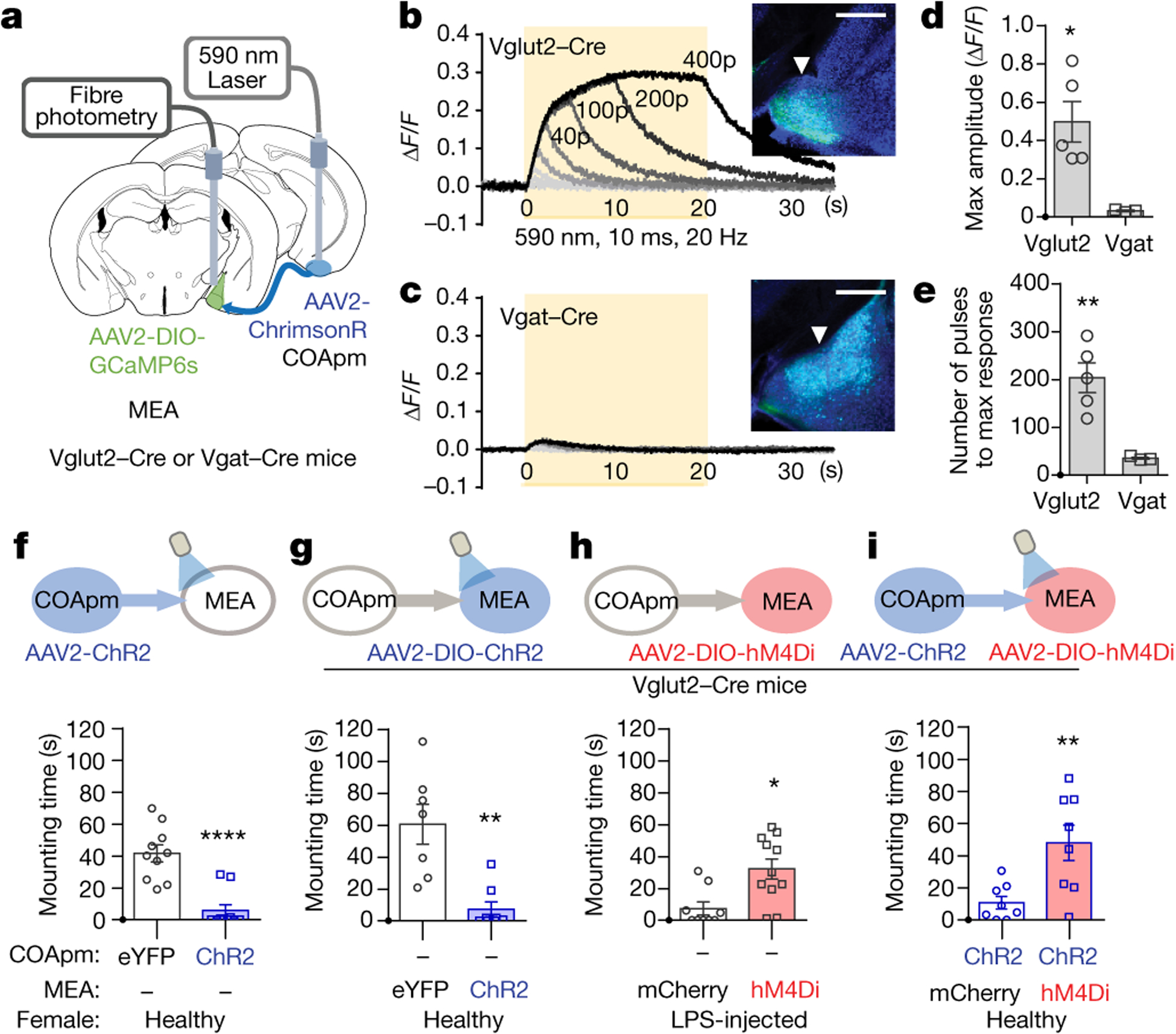
a-e, Red-shifted opsin ChrimsonR (AAV2-hSyn-ChrimsonR-tdTomato) was expressed in COApm, while GCaMP6s was expressed in MEA for fiber photometry analyses. Bulk fluorescence signal upon COApm photoactivation was measured in either MEA-Vglut2(+) or MEA-Vgat(+) neurons. Representative traces of changes in fluorescence signal in MEA upon photoactivation of COApm using light trains with 1, 5, 10, 20, 40, 100, 200 and 400 pulses, respectively. Inset images show GCaMP6s expression in MEA-Vglut2(+) or MEA-Vgat(+) neurons. Arrowheads indicate the placement of the optic fiber for fiber photometry (b,c). Maximum amplitude evoked by photoactivation of COApm with 400 pulses of light (d) and number of light pulses to reach max amplitude (e) (Vglut2-Cre, n=5 and Vgat-Cre, n=3; from 3 independent experiments). Scale bar=500μm. f-i, Quantification of male mounting behaviors during concurrent manipulation of COApm-MEA axonal projections and MEA-Vglut2(+) neurons. Male subjects were wild type for (f) or Vglut2-Cre mice for (g, h and i). Female subjects were healthy for (f, g and i) or LPS-injected for (h). f, Mounting time with photoactivation of COApm-MEA projections (EYFP, n=10 and ChR2, n=10; from 2 independent experiments). g, Mounting time with photoactivation of MEA-Vglut2(+) neurons (EYFP, n=7 and ChR2, n=8; from 2 independent experiments). h, Mounting time towards LPS-females with hM4Di-inhibition of MEA-Vglut2(+) neurons (mCherry, n=9 and hM4Di, n=11; from 2 independent experiments). i, Mounting time with concurrent photoactivation of COApm-MEA projections and hM4Di-inhibition of MEA-Vglut2(+) neurons (mCherry, n=8 and hM4Di, n=8; from 2 independent experiments). *P<0.05, **P<0.01 and ****P<0.0001 calculated by unpaired two-tailed t-test. Graphs indicate mean ± s.e.m. p-values are described in the supplementary statistical information file.