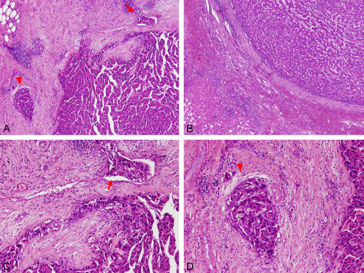
Figure 2.
Pathological features of the portal and capsular MVI. Sections after tumor resection were reviewed under microscopy by experienced pathologists. (A) Both capsule vein MVI (arrow) and portal vein MVI (arrowhead) under 40× magnification, (B) No capsular vein or portal vein MVI surrounding the tumor bed under 40× magnification, (C) Capsular vein MVI under 100× magnification: MVI (arrow) in surrounding tumor capsule (D) Portal vein MVI under 100× magnification: MVI in the portal vein with surrounding bile duct and hepatic arteriole.

