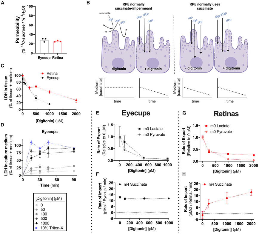
Figure 2. RPE-choroid permeability does not alter succinate uptake or oxidation.
(A) Permeability of freshly dissected retina and RPE tissue assessed by the relative uptake of 3H2O and 14C-sucrose over an hour ex vivo (n = 3).
(B) Model of the hypothesis to test whether succinate oxidation by RPE-choroid tissue is the result of cell permeability.
(C) Retina and RPE-choroid LDH release increases as a function of [digitonin]. Percentage of total was determined by assaying LDH in both tissue and culture medium when [LDH] was at a steady state in medium (n = 2–6 per concentration).
(D) Determination of the ex vivo culture time needed to reach steady-state [LDH] (n = 2–4 per concentration).
(E) Relative lactate and pyruvate release rate by RPE-choroid decreases with increasing [digitonin] (n = 8).
(F) RPE-choroid 13C4-succinate uptake rate however is unaltered by [digitonin] (n = 8).
(G) As with RPE-choroid tissue, lactate and pyruvate release decreases in retinas with increasing [digitonin] (n = 3–9).
(H) Unlike with RPE-choroid, retina 13C4-succinate uptake increases with [digitonin], suggesting that the plasma membrane is a barrier for retina but not RPE-choroid succinate uptake (n = 3–9). Data are represented as the mean ± SEM. (A) also shows values from individual replicates. See also Figure S2.

