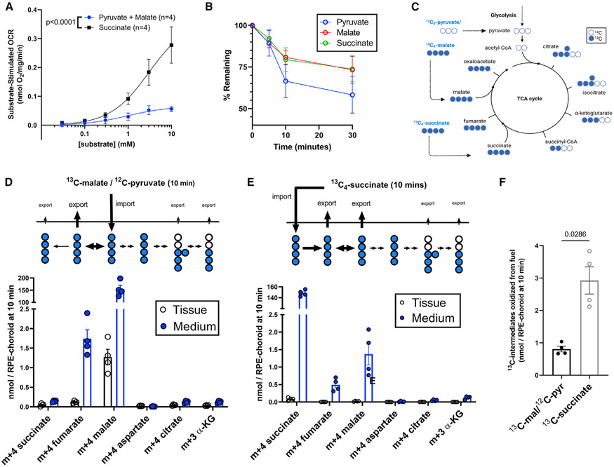
Figure 3. Succinate is preferred over pyruvate/malate for oxidation by RPE-choroid mitochondria.
(A) RPE-choroid OCR from 5 mM glucose supplemented with increasing concentrations (30 μM, 100 μM, 300 μM, 1 mM, 3 mM, 10 mM) of succinate or malate and pyruvate (n = 4).
(B) Pyruvate, malate, and succinate uptake (starting concentration: 1 mM ea.) by RPE-choroid ex vivo. Media was sampled before tissue was added; then 5, 10, and 30 min later, tissue was added (n = 7–8).
(C) TCA cycle 13C labeling scheme when cells use 13C4-malate/12C-pyruvate/12C-glucose or 13C4-succinate/12C-glucose as a substrate.
(D) Medium and tissue m+4 succinate, m+4 fumarate, m+4 malate, m+4 aspartate, m+4 citrate, and m+3 α-ketoglutarate in RPE-choroid incubated for 10 min in 1 mM 13C4-malate/1 mM 12C-pyruvate/5 mM 12C-glucose (n = 4).
(E) The same metabolites in medium and tissue of RPE-choroid incubated in 1 mM 13C4-succinate/5 mM 12C-glucose. In (C)–(D) the metabolite present in incubation medium was excluded from analysis (n = 4).
(F) Quantification of 13C in medium and tissue on energetically useful metabolites from each substrate, using data from (C)–(D). Reported p values result from (A) an extra sum-of-squares F test and (F) two-tailed Mann-Whitney tests. Data are represented as the mean ± SEM, with individual replicates also visible in (D)–(F). See also Figure S3.

