Fig. 4.

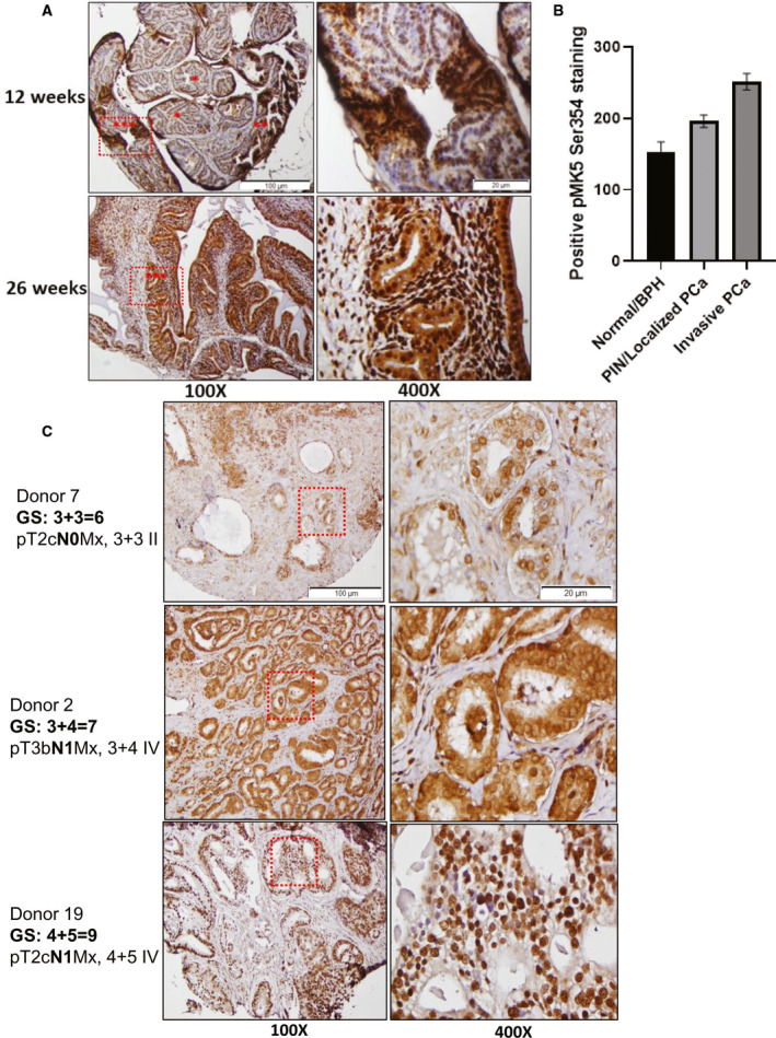
IHC staining revealed elevated pMK5 Ser354 level in PCa progression of TRAMP mice and in patients with high‐grade metastatic prostate. (A) Top panel, pMK5 Ser354 staining in a mixture of normal tissue/BPH, PIN/localized PCa and invasive PCa regions in 12‐week‐old mice. Bottom panel, pMK5 Ser354 staining in the invasive PCa lesions of 26‐week‐old mice that have spread throughout the gland. *= normal tissue/BPH, **= PIN/localized PCa and ***= invasive PCa. (B) ImageJ analysis of the positive staining of pMK5 Ser354 in different regions of TRAMP prostate tumour. Each figure is representative of three different TRAMP prostate tumours. One‐way ANOVA followed by Tukey’s post hoc analysis was used. Error bar represents standard error of the mean (SEM). (C) pMK5 Ser354 staining in TMA samples of Gleason scores 6, 7 and 9. Black dashed boxes represent the magnified regions. Scale bar: 100 µm (100×) and 20 µm (400×).
