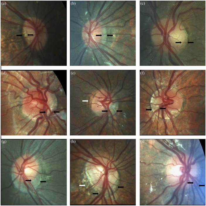
Figure 6.
Optic nerve crescents in children with Down syndrome. (a) Oval and tilted optic disk with a temporal crescent (black arrows) in a child with Down syndrome and myopia. (b) Choroidal crescent located temporally (black arrows) in a small, tilted disk from a child with Down syndrome and high myopia. (c) Small temporal crescent (black arrows) in a child with Down syndrome and hyperopia. (d) Small, tilted disk with vascular tortuosity. A scleral crescent is located below the disk and extends nasally (black arrows). (e) Tilted disk with situs inversus of the vessels (striped arrows). A large choroidal crescent is evident below the disk and extending into the nasal area (between the black arrows). Peripapillary atrophy is noted at the temporal margin of the disk (white arrows). (f) Tilted disk in which the scleral crescent, although wider below the disk, takes an annular form. Situs inversus, in which the vessels emerge nasally, is also evident (striped arrows). (g) Choroidal crescent, located below the disk with inferonasal and temporal extension (black arrows), in a child with Down syndrome and hyperopia. The disk appears equally tilted in this case. (h) Tilted and torted optic disk of a child with Down syndrome with myopic astigmatism. A choroidal crescent is evident below the disk (black arrows) along with a large zone of temporal peripapillary atrophy (white arrow). Note the bean-shaped optic disk in this case. (i) A smaller choroidal crescent, located below the disk and nasally, in a child with Down syndrome and hyperopia. In the upper and central rows, the optic disks have no physiological cupping. Reprinted from Postolache 27 2019 with permission from Frontiers.

