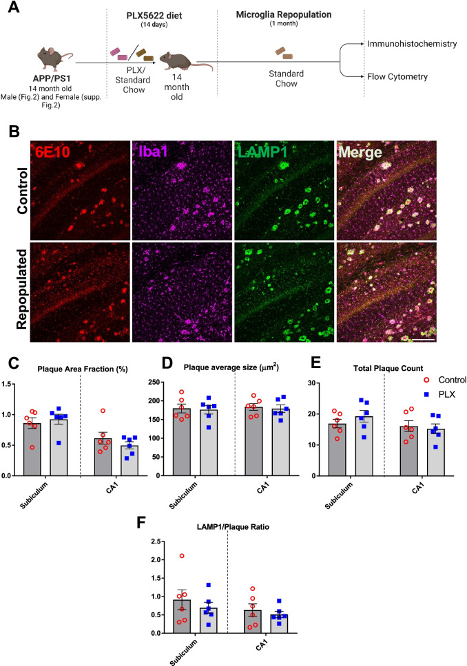
Fig. 2.
Repopulation of microglia does not ameliorate Aβ pathology and neuritic damage in APP/PS1 male mice. Experimental paradigm depicting duration of PLX5622 treatment and subsequent microglial repopulation (A). Representative immunofluorescent 20× images of the subiculum in control versus PLX-repopulated group, showing Aβ plaque (6E10, red), microglia (Iba1, magenta), and neuritic damage (LAMP1, green) (B). Scale bar represents 200 µm. There was no difference in the total area (C), size (D) and number (E) of plaques between the control and PLX-repopulated groups in Subiculum or CA1. Ratio of plaque-associated LAMP1+ neuritic damage to plaque load was similar between control and PLX-repopulated group (F). Student’s t-test. Data are presented as mean ± SEM (n = 6)

