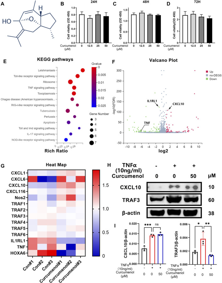
FIGURE 1.
Curcumenol showed little cytotoxicity in NP cells and down-regulated inflammatory pathways based on RNA-seq in vitro. (A) Chemical structure of Curcumenol. (B–D) Cell Counting Kit-8 assay results of NP cells stimulated with Curcumenol at different concentrations (0, 12.5, 25, and 50 μM) and different time periods (ranging from 24 to 72 h). (E) Ratio of up-regulated mRNA in NP cells treated with Curcumenol (50 μM) versus DMSO (1:1000) using KEGG pathway analyses (3 paired biological replicates). (F) Ratio of changed mRNA in NP cells treated with Curcumenol (50 μM) versus DMSO (1:1000) using Volcano Plot analyses (3 paired biological replicates). (G) Heat Map of changed mRNA in NP cells treated with Curcumenol (50 μM) versus DMSO (1:1000) using Volcano Plot analyses (3 paired biological replicates). (H) Western blot analysis of CXCL10 and TRAF3 expression in NP cells stimulated with TNFα (10 ng/ml) or/and 50 μM Curcumenol for 24 h (I) RT-qPCR analysis of the relative mRNA expression levels of CXCL10 and TRAF3 expression in NP cells stimulated with TNFα (10 ng/ml) or/and 50 μM Curcumenol for 24 h. All data are presented as mean ± SD from three replicates. *p < 0.05, **p < 0.01, ***p < 0.001 and ****p < 0.0001.

