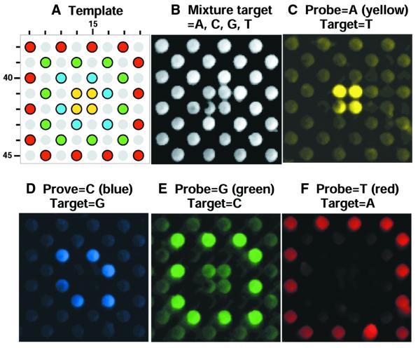
Figure 5.
FRE images of oDNA chip hybridization with fluorescein-labeled target sequences. A region of probes containing variation in a single-nucleotide position is shown (row and column coordinates are displayed). The site feature size is 150 µm. These hybridization experiments used the same oDNA chip. (A) The designed template of probes. The sites containing sequences with A, C, G or T in the variable position are color marked. Light gray circles are for probes of less than full length because of the deletion of 1–4 residues at certain residue positions in these sequences. (B) oDNA chip hybridization with a mixture of four target sequences which are complementary to all probe sequences in the region shown. (C–F) oDNA chip hybridization using a single-target sequence, which is complementary to the A, C, G or the T probe.

