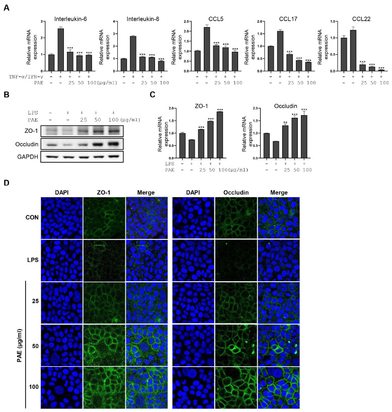
Fig. 3.
PAE suppresses inflammatory cytokine production and upregulates the tight junction in activated HaCaT cells. (A) The qRT-PCR analysis of IL-6, IL-8, CCL5, CCL17, and CCL22 is performed in TNF-α/IFN-γ-stimulated HaCaT cells after PAE treatment. (B, C) qRT-PCR and western blot analysis of ZO-1 and occludin are performed in LPS-stimulated HaCaT cells after dose-dependent PAE treatment. (D) Immunofluorescence microscopic analysis of ZO-1 and occludin are performed using LPS-stimulated HaCaT cells after dose-dependent PAE treatment. HaCaT cells were co-treated with PAE (100 μg/ml) and TNF-α/IFN-γ (10 ng/ml for each) for 24 hours. Data are expressed as means ± SD (n = 3). *P < 0.05; **P< 0.01; ***P< 0.001.

