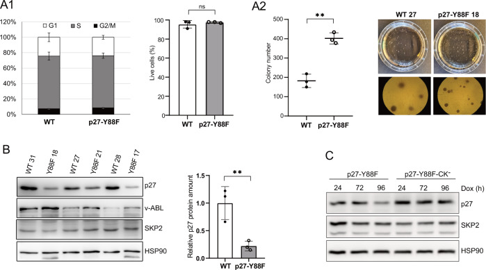
Fig. 6. Colony formation capacity of established p27-Y88F v-ABL+ cell lines is increased and downregulation of p27-Y88F depends on cyclin-CDK binding.
A1 Left graph represents cell cycle phase distribution of v-ABL+ wild type and p27-Y88F cells as determined by flow cytometry after propidium iodide and anti-BrdU staining. Right graph indicates percentages of live cells identified by flow cytometry as cells staining negative for Annexin V and DAPI. A2 Colony formation assay of wild type and p27-Y88F v-ABL+ cells. Left panel displays colony numbers of one representative experiment out of two. Right panel shows photographs of a representative wild type and p27-Y88F colony formation assay. B Cells isolated from colonies were subjected to western blot. p27, v-ABL and SKP2 protein levels were analyzed. HSP90 was used as loading control. Right panel presents densitometric analysis of p27 protein levels normalized to HSP90. Data represent mean ± SD. (**p < 0.01). C p27-Y88F or p27-Y88F-CK− expression was induced with 1 μg/ml doxycycline in HeLa Flp-In cells for 24, 72 and 96 h and p27, SKP2 and HSP90 protein levels were analyzed by western blot. One representative experiment out of three is shown.

