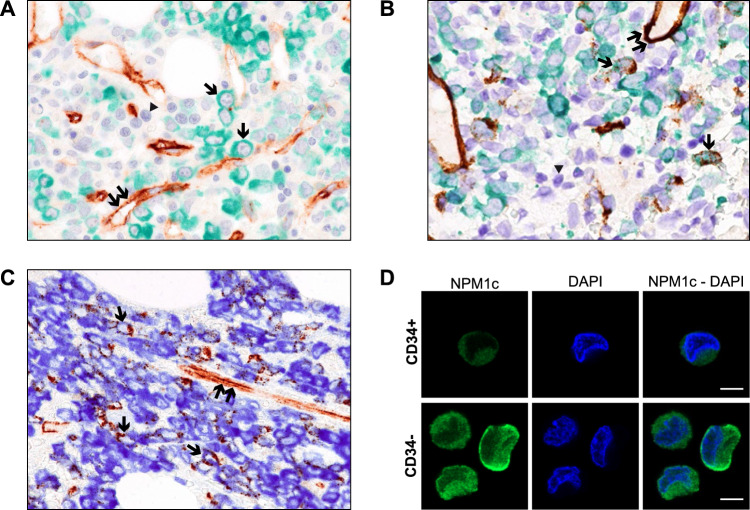
Fig. 2. Co-expression of NPM1 mutant and CD34+ in NPM1-mutated AML.
A. Formalin-fixed BM paraffin section from NPM1-mutated AML at diagnosis double stained for CD34 (brown, peroxidase with DAB chromogen) and mutant NPM1 protein (green, peroxidase with green chromogen, DC9913, Leica) plus BOND Polymer Refine HRP PLEX Detection and counterstaining in hematoxylin (blue). Single arrows point to leukemic cells expressing cytoplasmic NPM1 (green) but not CD34. Cells stained only in blue (hematoxylin) represent normal residual hematopoietic cells that do not express CD34 and the NPM1 mutant protein (arrowhead). Double arrows indicate CD34+ vessel endothelial cells (brown) that serve as positive control (magnification, x400). B Formalin-fixed BM paraffin section from another NPM1-mutated AML sample at diagnosis double stained with CD34 (brown) and mutant NPM1 (green) and counterstained in hematoxylin (blue) as indicated in panel A. Most leukemic cells show cytoplasmic NPM1 (green) in the absence of CD34 (brown) whilst a few blast cells (single arrows) are double stained (brown/green) for CD34 and cytoplasmic NPM1 mutant protein. Cells stained only in blue (hematoxylin) represent normal residual hematopoietic cells that do not express CD34 and the NPM1 mutant protein (arrowhead). Double arrows indicate CD34+ vessel endothelial cells (brown) that serve as positive control (magnification, x400). C Formalin-fixed BM paraffin section from a NPM1-mutated AML patient at relapse double stained for CD34 (brown, peroxidase with DAB chromogen) and mutant NPM1 protein (blue, peroxidase with blue chromogen, DC9896, Leica) plus BOND Polymer Refine Detection, without hematoxylin counterstaining. A significant percentage of leukemic cells (single arrows) are double stained (brown/blue) for CD34 and NPM1 mutant protein. Double arrows indicate CD34+ vessel endothelial cells (brown) that serve as positive control (magnification, x400). D Immunofluorescence with anti-NPM1 mutant antibody (green) on the CD34+ and CD34− sorted cells. Nuclei were stained with DAPI (blue). 63x magnification. Scale bar, 5 μm.

