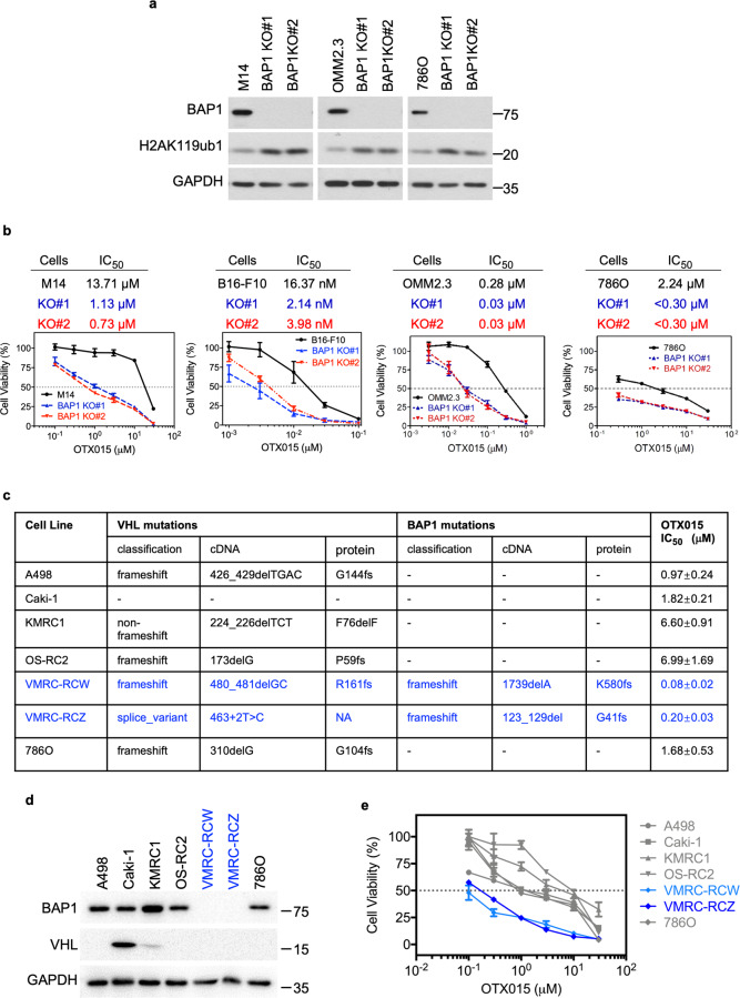
Fig. 2. BAP1 loss augments sensitivity to BET inhibitors.
a Immunoblot analysis showing the deletion of BAP1 and increased H2AK119ub1 in M14, OMM2.3, and 786O cells. Two clones for each cell line (#1 and #2) are shown. b The BET inhibitor OTX015 exhibited potent and selective cytotoxicity against BAP1-deficient M14, B16-F10, OMM2.3, and 786O cells. Cells were treated with OTX015 at the indicated concentrations for 6 days. The data are presented as the mean ± SD (n = 3) from one representative experiment out of three. c The mutation status of VHL and BAP1 as well as OTX015 IC50 values are shown for each ccRCC cell line. The mutation data are from Cell Model Passport Databases (http://cellmodelpassports.sanger.ac.uk). The IC50 values for each cell line are shown as the mean ± SD (n = 3) from three independent experiments. d Immunoblot analysis showing VHL and BAP1 expression in multiple ccRCC cell lines. e Cell viability of multiple ccRCC cell lines with wt or mutant BAP1 in response to OTX015 treatment at the indicated concentrations for 6 days. The data are presented as the mean ± SD (n = 3) from one representative experiment out of three.

