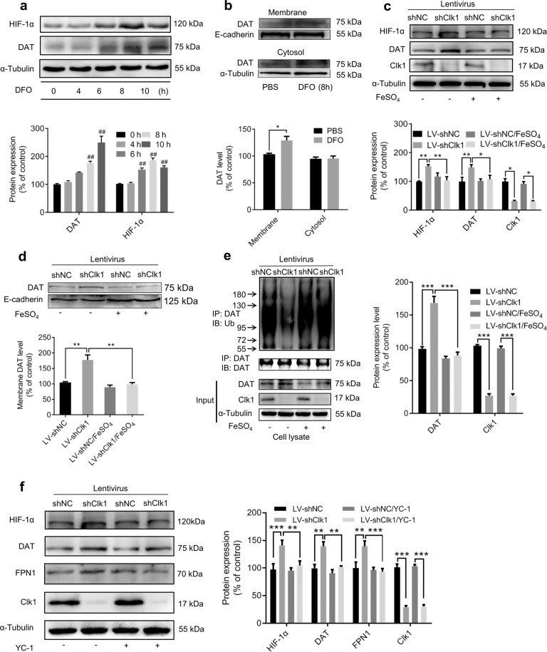
Fig. 6. HIF-1α modulates iron deficiency-induced plasma membrane DAT expression.
a PC12 cells were treated with 100 μM DFO for indicated periods prior to immunoblot assay and b membrane protein of PC12 cells was isolated as described in Methods. Cell lysates were prepared for immunoblotting against specific antibodies. c, d LV-shNC and LV-shClk1 PC12 cells were treated with 100 μM FeSO4 for 8 h prior to immunoblot assays and e DAT-ubiquitin assay. f YC-1 (100 μM) was added to LV-shNC and LV-shClk1 PC12 cells for 24 h prior to immunoblot assays. Bar graphs are presented as means ± SEM for at least 3 independent experiments. For each of a–d, representative images for immunoblots are shown in the upper panels and quantitative data are shown in the lower panels. For e and f, representative images for immunoblots are shown in the left panels and quantitative data are shown in the right panels. Statistical analyses for a–c, e and f were performed using two-way ANOVA and statistical analysis for d was performed using one-way ANOVA, each followed by Bonferroni-corrected tests (*P < 0.05, **P < 0.01, ***P < 0.001; ##P < 0.01 vs 0 h).

