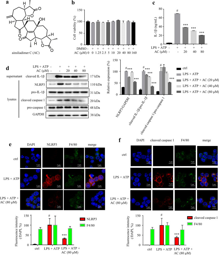
Fig. 1. AC suppressed LPS plus ATP-stimulated IL-1β secretion in THP-1 cells.
a Chemical structure of AC. b Cytotoxicity of AC on THP-1 cells assessed by MTT assay. THP-1 cells were treated with different concentrations of AC (from 1.25 to 160 μM) for 24 h (n = 6). c The levels of IL-1β in the culture medium from THP-1 cells (n = 3). THP-1 cells were cultured in the presence or absence of AC for 12 h, and stimulated with LPS for 4 h and then ATP for 1 h. d The expression of NLRP3, pro-caspase 1, cleaved caspase 1, pro-IL-1β in the lysates, and cleaved IL-1β in the supernatant of THP-1 cells detected by Western blotting. GAPDH was chosen as an internal loading control (n = 5). Immunofluorescence staining of NLRP3 (e) and cleaved caspase 1 (f) (n = 3). Scale bar = 10 μm. Data are expressed as means ± SEM. #P < 0.05 versus DMSO, ***P < 0.001 versus LPS + ATP.

