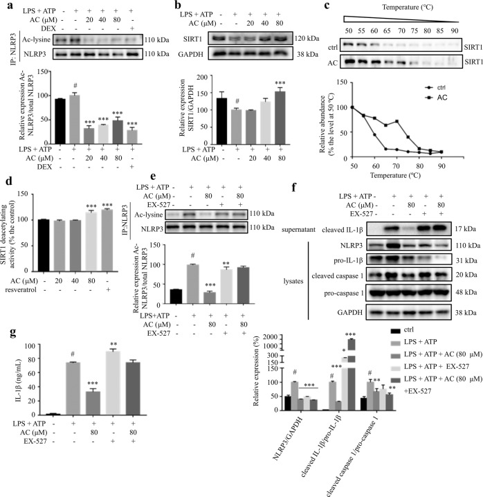
Fig. 2. AC inhibited the activation of NLRP3 inflammasome through activating SIRT1.
THP-1 cells were treated with various concentrations of AC for 12 h, and then stimulated with LPS for 4 h and ATP for 1 h. a The acetylated and total NLRP3 protein levels (n = 3). b The expression level of SIRT1 detected by Western blotting (n = 3). GAPDH was chosen as an internal loading control. c CETSA performed on THP-1 cells after the treatment with or without AC (80 μM) for 12 h (n = 3). d The effect of AC or resveratrol on the deacetylating activity of SIRT1 (n = 6). The deacetylating activity was determined with a SIRT1 fluorometric assay kit. e The acetylated and total NLRP3 protein levels (n = 3). f The expression of NLRP3, pro-caspase 1, cleaved caspase 1, and pro-IL-1β in the lysates, and cleaved IL-1β in the supernatant of THP-1 cells detected by Western blotting (n = 3). GAPDH was chosen as an internal loading control. g The levels of IL-1β in the culture medium from THP-1 cells (n = 6). Data are expressed as means ± SEM. #P < 0.05 versus DMSO, **P < 0.01, ***P < 0.001 versus LPS + ATP.

