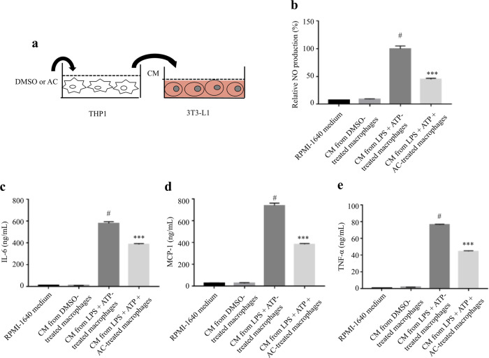
Fig. 5. AC attenuated macrophage-CM-induced inflammatory responses in adipocytes.
a Schematic diagram of the experimental procedure. THP-1 cells were treated AC (80 μM) or DMSO for 12 h, and subsequently stimulated with LPS for 4 h and ATP (1 mM) for 1 h. After 24 h of culturing in fresh medium, the supernatants were collected as macrophage CM, which was used to incubate the fully differentiated 3T3-L1 for 24 h. The production of NO (b) and the levels of IL-6 (c), MCP-1 (d), and TNF-α (e) in 3T3-L1 adipocytes were determined. Data are expressed as means ± SEM. n = 6. #P < 0.05 versus DMSO, ***P < 0.001 versus LPS + ATP.

