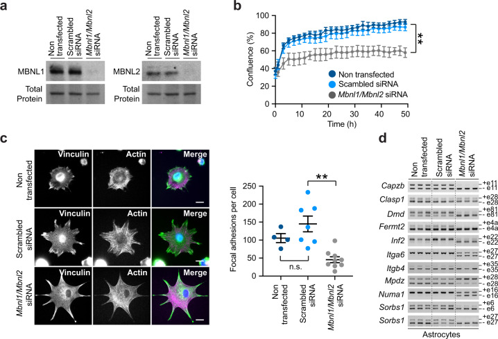
Fig. 8. MBNL1 and MBNL2 inactivation affects astrocyte cell growth, adhesion, and alternative splicing.
a Western blot detection of MBNL1 and MBNL2 in double knockdown primary astrocytes, relative to scramble siRNA and non-treated controls. Representative stain-free protein bands are shown to illustrate total protein loading. The experiment was performed on two independent cultures per treatment. b Monitoring of the confluence of primary astrocytes by live cell video-miscroscopy, from 45 min after plating up to 48 h. Primary MBNL1/MBNL2 knockdown astrocytes are compared with non-transfected and scrambled-transfected controls. Data are means ± SEM, n = 4 independent cultures per genotype (p = 0.0089, Two-way repeated measures ANOVA). c Immunofluorescent quantification of vinculin-rich focal adhesions per individual cell in siRNA-treated primary astrocytes, following 3 h in culture. Scale bar, 20μm. Data are means ± SEM. N = 2 independent cultures per cell group; n = 4 cells, non-transfected; n = 7 cells, scrambled siRNA; n = 9 cells, Mbnl1/Mbnl2 siRNA (p > 0.9999, non-transfected vs scrambled siRNA; p = 0.0023, scrambled siRNA vs Mbnl1/Mbnl2 siRNA; Kruskal–Wallis test, Dunn’s post hoc test for multiple comparisons). d RT-PCR splicing analysis of adhesion- and cytoskeleton-associated transcripts in Mbnl1/Mbnl2 double knockdown astrocytes, compared with non-transfected and scrambled siRNA controls. Alternative exons are shown on the right. The experiment was performed on three independent cultures per treatment. Source data are provided as a Source Data file. n.s. not significant; **p < 0.01.

