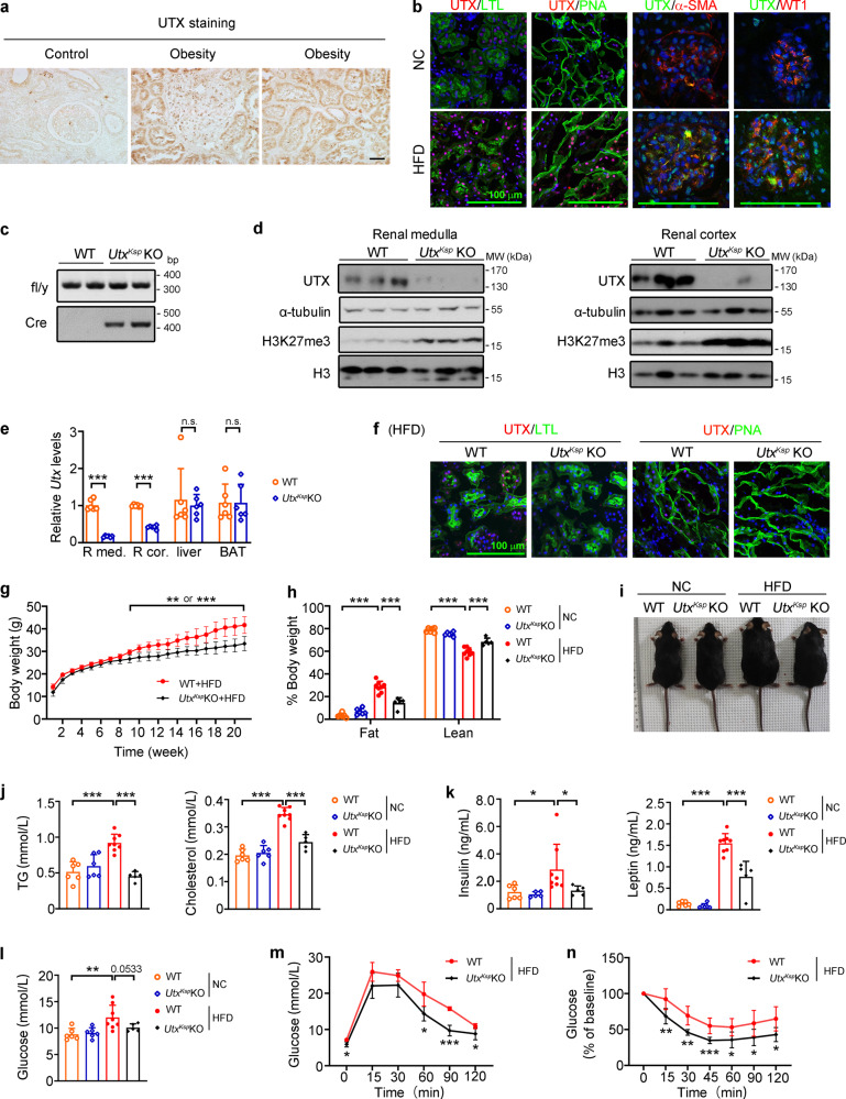
Fig. 1. UtxKsp KO mouse was resistant to HFD-induced obesity.
a Representative images of renal UTX staining of human subjects, n = 4 or five independent subjects with control or obesity, respectively. Scale bar, 50 μm. Brown color indicates positive staining. b Representative images of UTX (red)/LTL (lotus tetragonolobus lectin, detecting proximal tubules; green), UTX (red)/PNA (peanut agglutinin, detecting distal tubules and collecting ducts; green), UTX (green)/α-SMA (alpha-smooth muscle actin, mesangial cell marker; red), and UTX (green)/WT1 (Wilms’ tumor 1, podocyte marker; red) staining on the renal sections of WT (n = 6) and HFD (n = 4) mice. DAPI staining shown in blue. Scale bar, 100 μm. c Genotyping of WT and UtxKsp KO mice. d Western blots of UTX, α-tubulin, H3K27me3 and H3 in renal medulla (left, n = 4) and cortex (right, n = 6) of WT and UtxKsp KO mice. e qPCR results of Utx in renal medulla (R med.), renal cortex (R cor.), liver and BAT of WT and UtxKsp KO mice. n = 6 independent animals (mean ± SD); ***PR med. < 0.0001, ***PR cor. < 0.0001 (unpaired, two-tailed t-test). f Representative images of UTX (red)/LTL (green), UTX (red)/PNA (green) staining on the renal sections of HFD-fed WT and UtxKsp KO mice. DAPI (blue) stained nuclei. Scale bar, 100 μm, n = 4 independent animals. g–i, Growth curves (g), body mass (h), and gross view (i) of WT and UtxKsp KO mice. g n = 8, 5 independent animals for WT + HFD or UtxKsp KO + HFD group, respectively (mean ± SD), **Pbody weight or ***Pbody weight from 0.0035 to 0.0007; unpaired, two-tailed t-test; h n = 6, 6, 8, five independent animals for WT + NC or UtxKsp KO + NC or WT + HFD or UtxKsp KO + HFD group, respectively (mean ± SD), ***PFat (WT+NC vs WT+HFD) < 0.0001, ***PFat (WT+HFD vs UtxKsp KO+HFD) < 0.0001; ***PLean (WT+NC vs WT+HFD) < 0.0001, ***PLean (WT+HFD vs UtxKsp KO +HFD) < 0.0001 (one-way ANOVA). j–l Serum levels of TG (j, left), cholesterol (j, right), insulin (k, left), leptin (k, right) and non-fasting blood glucose (l) of WT and UtxKsp KO mice, n = 6, 6, 8, 5 independent animals for WT + NC or UtxKsp KO + NC or WT + HFD or UtxKsp KO + HFD group, respectively (mean ± SD); ***PTG (WT+NC vs WT+HFD) < 0.0001, ***PTG (WT+HFD vs UtxKsp KO+HFD) < 0.0001; ***Pcholesterol (WT+NC vs WT+HFD) < 0.0001, ***Pcholesterol (WT+HFD vs UtxKsp KO+HFD) < 0.0001; *PInsulin (WT+NC vs WT+HFD) = 0.0294, *PInsulin (WT+HFD vs UtxKsp KO+HFD) = 0.0378; ***PLeptin (WT+NC vs WT+HFD) < 0.0001, ***PLeptin (WT+HFD vs UtxKsp KO+HFD) < 0.0001; **Pglucose (WT+NC vs WT+HFD) = 0.0027 (one-way ANOVA). m, n GTT (m) and ITT (n) results of WT and UtxKsp KO mice. m n = 4, five independent animals for WT + HFD or UtxKsp KO + HFD group, respectively (mean ± SD), *PGTT(0) = 0.0214; *P GTT(60) = 0.0373; ***P GTT(90) = 0.0003, *PGTT(120) = 0.0428; (n) n = 8, five independent animals for WT + HFD or UtxKsp KO + HFD group, respectively (mean ± SD), **PITT(15) = 0.0063, **PITT(30) = 0.0011, ***P ITT(45) = 0.0009, *P ITT(60) = 0.0238, *P ITT(90) = 0.0341, *P ITT(120) = 0.0125, unpaired, two-tailed t-test. Source data are provided in the Source Data file.

