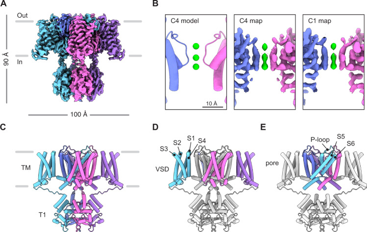
Fig. 1. Structure of human Kv1.3.
A Cryo-EM density map of human Kv1.3. B The pore region of Kv1.3 as modeled with C4 symmetry (left), as seen in the density map refined with C4 symmetry (middle) or as modeled with C1 symmetry (right). This panel highlights the presence of three K+ ion densities and the equivalent ion occupancy when using C1 and C4 symmetries. K+ ions shown in green. C Model of human Kv1.3 with C4 symmetry constraints. The transmembrane (TM) region and T1 domains are indicated. Model of Kv1.3 with the voltage sensing domains (VSDs) colored (D), or with the pore domain colored (E). The front and rear VSDs are omitted for clarity in E.

