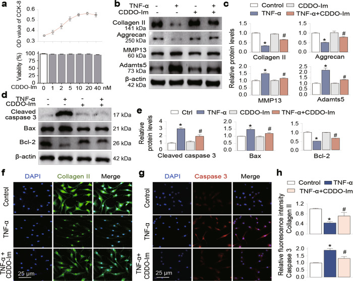
Fig. 1. CDDO-Im attenuates TNF-α-induced chondrocyte apoptosis and ECM degradation.
a Cell viability assay. Primary chondrocytes were treated with various doses of CDDO-Im for 24 h. Cell viability was assayed by a cell counting kit (CCK-8, upper panel) and the trypan blue exclusion test (lower panel). Western blot analysis of protein expression and quantification of Collagen II, Aggrecan, MMP13 and Adamts5 (b and c) and cleaved caspase 3, Bax and Bcl-2 (d and e) in chondrocytes treated with CDDO-Im (20 nM) with or without TNF-α (50 ng/mL) for 24 h. Representative images of immunofluorescence staining of Collagen II (f) or Caspase 3 (g). Chondrocytes were treated with CDDO-Im (20 nM) with or without TNF-α (50 ng/mL) for 24 h. h Quantitative analysis of the data in Fig. 1f and g. The values are presented as the means ± SDs. *P < 0.05 versus control, #P < 0.05 versus TNF-α-treated chondrocytes.

