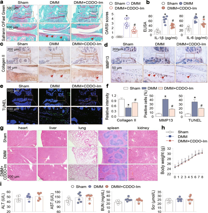
Fig. 2. CDDO-Im ameliorates DMM-induced cartilage erosion in mouse knee joints, chondrocyte apoptosis and OA pathologies.
Mice were divided into Sham, DMM, and CDDO-Im-treated DMM groups (n = 6 per group, 8 weeks). a Representative images of safranin-O/fast green stained knee joint sections from sham, DMM and CDDO-Im-treated DMM mice. The arrows indicated the damaged cartilage areas. OARSI scores were on the right side. b The serum levels of IL-1β and IL-6 were measured by ELISA. c Representative immunohistochemical (IHC) staining of Collagen II and d MMP13. The arrows indicate damaged cartilage areas (c) or positively- stained chondrocytes (d). e Representative images of TUNEL staining of the sections. f Quantitative analysis of the data in Fig. 2c, d and e. g Representative H&E staining images of heart, liver, lung, spleen and kidney of sham, DMM and CDDO-Im-treated DMM mice. h Body weight changes of sham, DMM and CDDO-Im-treated DMM mice during the 8-week of experimental period. i Serum levels of alanine aminotransferase (ALT), aspartate aminotransferase (AST), blood urea nitrogen (BUN) and serum creatinine (Scr). The values are presented as means ± SEMs. *P < 0.05 versus sham mice, #P < 0.05 versus DMM mice.

