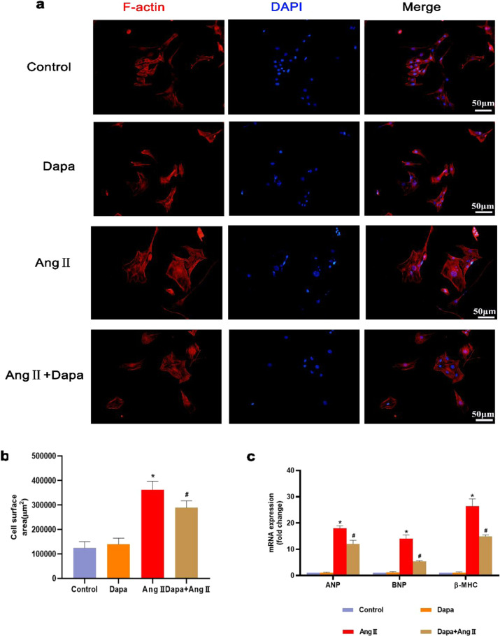
Fig. 3. DAPA attenuated Ang II-induced cardiac hypertrophy in vitro.
a The cell surface area of HL-1 cells was assessed by TRITC-conjugated phalloidin staining. b Calculated cell surface area (>100 cells per group). c ANP, BNP and β-MHC mRNA levels were assessed by real-time PCR. n = 6, ∗P < 0.05 versus the CON or DAPA group, #P < 005 versus the Ang II.

