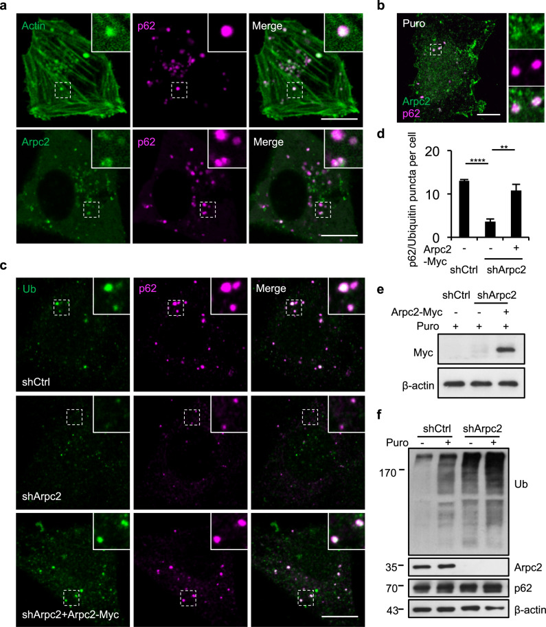
Fig. 2. The Arp2/3 complex is essential for the formation of p62 bodies.
a Cells were transfected with GFP-Actin or Arpc2-GFP together with tdTomato-p62, and then imaged. Regions containing p62 foci that co-localize with branched actin filaments are outlined with white dashed lines and are magnified in the insets. Scale bars, 10 µm. b Cells were treated with 1 mg/mL puromycin for 8 h and stained with antibodies against Arpc2 and p62. Scale bar, 10 µm. c NRK cells expressing non-targeting control shRNA (shCtrl), Arpc2 shRNA, or Arpc2 shRNA plus shRNA-resistant Arpc2-Myc cDNA were treated with 1 mg/mL puromycin for 8 h, and then stained with ubiquitin and anti-p62 antibodies. Scale bar, 10 µm. d The number of puncta positive for both ubiquitin and p62 was quantified in cells from c (n = 3 independent experiments; 50 cells were assessed per independent experiment). P-values were calculated using the two-tailed unpaired t-test. **P < 0.01; ****P < 0.0001. e Immunoblot showing the protein expression level in NRK cells with ectopic expression of Myc-tagged Arpc2. f NRK cells expressing shCtrl or Arpc2 shRNA were treated with 1 mg/mL puromycin for 8 h, and the cell lysates were analyzed by western blotting with antibodies against ubiquitin, Arpc2, β-actin, and p62.

