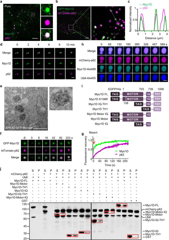
Fig. 3. Myosin 1D coalesces with p62 bodies.
a NRK cells were treated with 1 mg/mL puromycin for 8 h, and stained with antibodies against Myo1D and p62. Scale bar, 10 μm. b NRK cells were transfected with GFP-Myo1D and tdTomato-p62, and observed by SIM. Scale bar, 10 µm. c Line profiling of a representative section of the cell, indicated by the white dashed line in b. d Time-lapse images of p62 bodies in live ATG12 KO NRK cells transiently expressing GFP-Myo1D and tdTomato-p62. Scale bar, 1 µm. e TEM image showing the DAB staining pattern in NRK cells transiently transfected with APEX2-GFP-Myo1D and tdTomato-p62. Scale bar, 1 µm. f Fluorescence intensity recovery of a p62 body in an NRK cell transiently expressing GFP-Myo1D and tdTomato-p62 after photobleaching. Scale bar, 1 µm. g Quantification of fluorescence intensity recovery of a photobleached p62 body (n = 3). h Droplets formed by LLPS of p62 (10 μM), Ub8 (6 μM) and Myo1D (1 μM) fusion upon contact in vitro. Scale bar, 5 μm. i Schematic diagram of Myo1D constructs. EGFP was fused to the N-terminus of FL Myo1D, Myo1D K108R, Myo1D IQ-TH1, Myo1D TH1, Myo1D Motor-IQ or Myo1D IQ. j Representative sedimentation analysis showing Myo1D fragments concentrated in the soluble phase (S) or condensed phase (P). Results were analyzed by western blotting using anti-His tag antibody.

