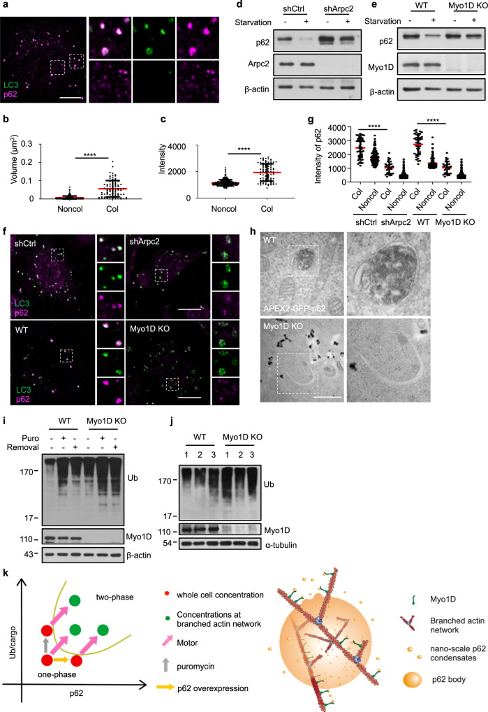
Fig. 5. The branched actin network regulates autophagy by harnessing p62 phase condensation.
a NRK cells were starved for 4 h, and then stained with antibodies against LC3 and p62. The left panel shows the colocalization of p62 and LC3. Right panels show enlarged p62 structures that were either LC3-positive (upper panels) or LC3-negative (lower panels). Scale bar, 10 µm. b, c Size (b) or intensity (c) variation of p62 bodies that are colocalized (Col) or non-colocalized (Noncol) with LC3 in a. Size was quantified as volume (μm3). P-values were calculated using the two-tailed unpaired t-test (n = 3). ****P < 0.0001. d Cells transfected with non-targeting control shRNA (shCtrl) or Arpc2 KD shRNA (shArpc2) were starved with DPBS (STA) for 2 h, and then the cell lysates were analyzed by western blotting with antibodies against p62 and β-actin. e WT cells or Myo1D KO cells were starved with DPBS (STA) for 2 h, and then the cell lysates were analyzed by western blotting with antibodies against p62 and β-actin. f Cells transfected with shCtrl or shArpc2 or Myo1D KO cells were starved for 4 h, and then stained with antibodies against p62 and LC3 followed by confocal microscopy analysis. Right panels show enlarged p62 structures which were LC3 positive. Scale bar, 10 µm. g Intensity variation of p62 bodies that are colocalized or non-colocalized with LC3 in each group in f after nutrient starvation. P-values were calculated using the two-tailed unpaired t-test (n = 3). *P < 0.05; ****P < 0.0001. h TEM images showing the DAB staining pattern in WT NRK cells and Myo1D KO cells transiently expressing APEX2-GFP-p62 followed by starvation for 4 h. Scale bar, 1 μm. i WT and Myo1D KO NRK cells were treated with 1 mg/mL puromycin for 8 h. After puromycin washout, the cells were cultured in normal conditions for 16 h, and then the cell lysates were analyzed by western blotting with antibodies against ubiquitin and Myo1D. j Brain lysates of WT and Myo1D KO mice were analyzed by western blotting with antibodies against ubiquitin and Myo1D. k Left, phase diagram as a function of the branched actin networks and motor proteins for condensate formation. Right, a working model showing that the branched actin network and Myo1D-mediated active transportation drive the condensation of p62 bodies.

