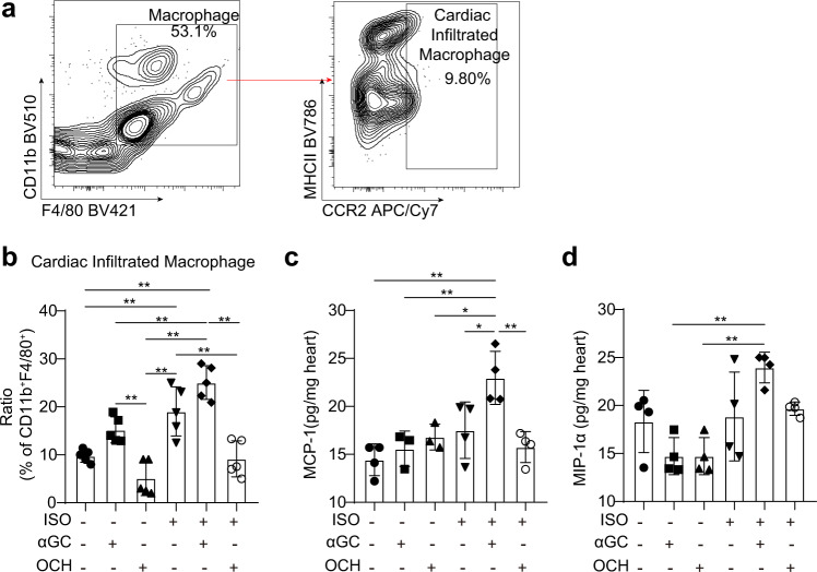
Fig. 8. Cardiac macrophages in αGC-induced cardiotoxicity.
a–d Hearts were harvested from mice stimulated with ISO (5 mg/kg per day) alone for 7 days or together with αGC or OCH every other day. a FACS was used to analyze the proportions of macrophages and adult monocyte-derived macrophages (infiltrated) in the hearts. b The percentage of CCR2-positive macrophages among CD11b+ F4/80+ cells was calculated. c, d Whole-heart lysates were subjected to ELISA immunoassays to determine the protein concentrations of MCP-1 and MIP-1α. The data are expressed as the mean ± SD. *P < 0.05 and **P < 0.01.

