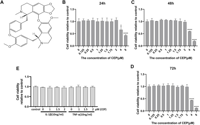
FIGURE 1.
Effects of CEP on chondrocyte viability. (A) Molecular structure of CEP. (B,C,D) Cells were treated with the indicated concentrations of CEP for 24, 48, and 72 h. Mouse chondrocyte viability was evaluated by CCK-8 assay. (E) Chondrocytes were exposed to 10 ng/ml IL-1β or TNF-α with or without CEP (1, 1.5, and 2 μM), and cell viability was determined by CCK-8 assay. All experiments were repeated independently three times. Values are expressed as mean ± SD; *p < 0.05 and ****p < 0.0001 vs. the control group.

