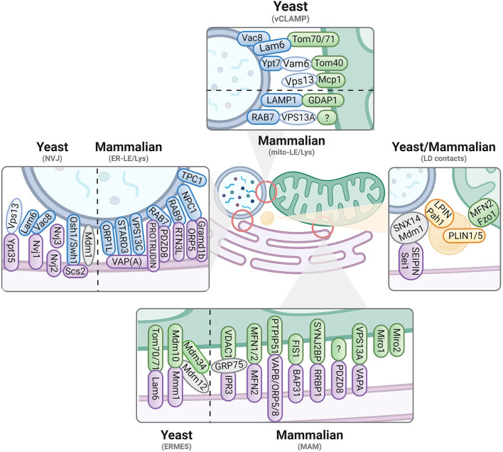
Figure 1.
Contact sites in yeast and mammalian cells. Known tethers and interactors are displayed for contacts involving the ER (magenta), mitochondria (green), vacuoles/endolysosomes (blue) and lipid droplets (yellow). Yeast versus mammalian interactors are separated where possible. Proteins identified in yeast are written in lowercase, while mammalian/human proteins are in capital letters. For more details, we refer to Table 1.

