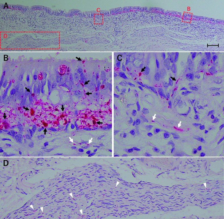
Fig. 4.
Microscopic images of OE with fibrous changes in the septum area of the olfactory clefts, immunostained for phosphorylated tau (AT8) (A). Some degenerated olfactory cells (black arrows) and their axons (white arrows) in the lamina propria of this epithelium stained positive for phosphorylated tau. Olfactory fascicles also stained partially positive for phosphorylated tau (white arrowheads) (D). Bar = 100 μm.

