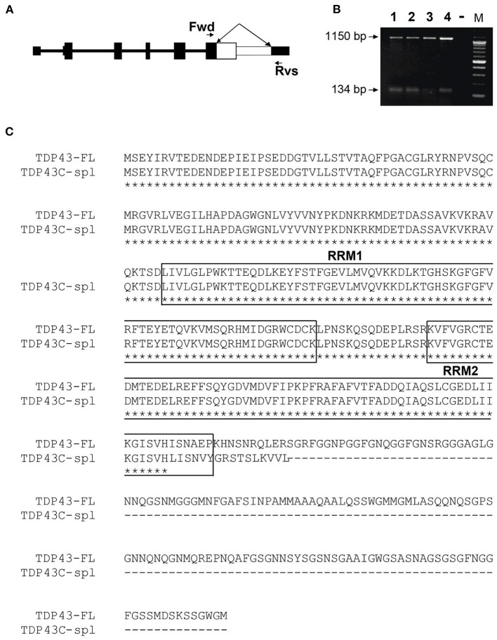
Figure 2.
Identification of a human TDP-43 splice variant using RT-PCR. (A) Representation of the gene structure of full length TDP-43. The forward (Fwd) primer in CDS of exon 6 and reverse (Rvs) primer in the 3′UTR in exon 6 are indicated by arrows. The white area in exon 6, indicated by the double-headed arrow, represents the region that is spliced out in TDP43C-spl. (B) A major higher RT-PCR product of 1,150 bp, corresponding to the human full-length TDP-43 (TDP43-FL), was amplified from RNA isolated from Lane 1, human brain; Lane 2, human spinal cord; Lane 3, human dorsal root ganglion and Lane 4, SHSY5Y cells. The lower band represents a cluster of exon 6 splice variants, with DNA sequencing identifying TDP43C-spl, with a 1,020 bp deletion generating a RT-PCR product of 134 bp. (C) Sequence alignment of TDP43-FL and TDP43C-spl. The two RNA-recognition motifs (RRM1 and RRM2) in TDP43-FL are mostly conserved in TDP43C-spl (boxed). A unique 16 aa sequence (LISNVYGRSTSLKVVL) is generated at the C-terminus of TDP43C-spl.

