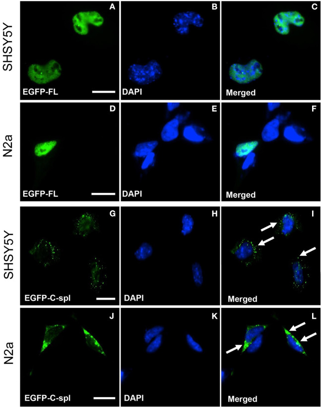
Figure 4.
Subcellular localization of TDP43-FL and TDP43C-spl in neuronal cells. SHSY5Y (human neuroblastoma) and N2a (mouse neuroblastoma) cells were transfected with EGFP-tagged TDP43-FL (A–F) or EGFP-tagged TDP43C-spl (G–L). The EGFP-tagged TDP43-FL (A,D) and blue DAPI nuclear stain (B,E) showed co-localization to the nucleus in both SHSY5Y cells (C) and N2a cells (F). EGFP-tagged TDP43C-spl was localized to the cytoplasm in both SHSY5Y cells (G) and N2a cells (J), with no co-localization with DAPI [(H,K), with overlays shown in (I,L)]. Cytoplasmic localization of aggregate TDP43C-spl is shown by arrows in (I,L). Scale bars = 15 μm.

