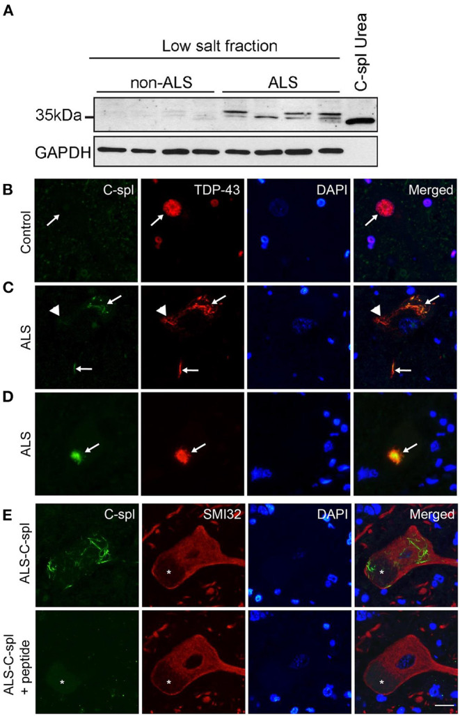
Figure 8.

TDP43C-spl/sTDP-43 isoforms are incorporated into TDP-43 positive pathological inclusions in ALS motor neurons. (A) Lysates of non-ALS (n = 4) and ALS (n = 4) lumbar spinal cord show increased expression of TDP43C-spl/sTDP43 in ALS cases compared to controls. GAPDH is used as a loading control and lysate of N2a cells transiently transfected with TDP43C-spl was used as a positive control. Double immunofluorescence staining of spinal cord motor neurons of (B) control and (C–E) ALS cases with CTUS antibody (green); TDP43-FL (red); and DAPI stain. (B) In control cases, there was no apparent CTUS staining in the nucleus (arrows). Note that in (C,D) there was co-labeling of CTUS antibody with TDP43-FL in (C) skein-like inclusions (arrows), and (D) round inclusions (arrows), however this co-labeling was only partial (arrowheads). (E) Specificity of labeling was confirmed by competition with the immunizing peptide on serial sections. Motor neurons were identified using non-phosphorylated neurofilament antibody (SMI32, red). Asterisks indicate lipofuscin. Scale bar = 15 μm.
