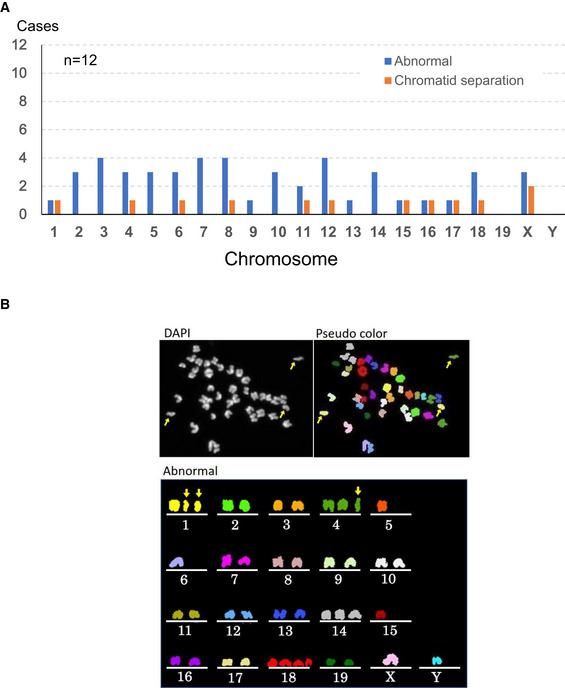
Figure EV3. Chromosomal multicolor FISH analysis of MII oocytes derived from spermatocyte injection.

- Chromosomal abnormalities were found in both autosomes and sex chromosomes.
- A representative image of an oocyte with chromosomal aberrations. Arrows indicate prematurely separated chromatids. Besides them, chromosomes 5, 6, 14, 15 and 18 were numerically abnormal.
