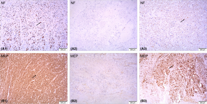
FIGURE 3.

Immunohistochemical staining of spinal cord tissue removed during the surgery. NF‐200‐positive axons and MBP‐positive myelin sheaths can be observed at the distal and proximal ends of the tissue (arrows in A1, B1, A3, B3) but not in the center of the tissue (A2, B2)
