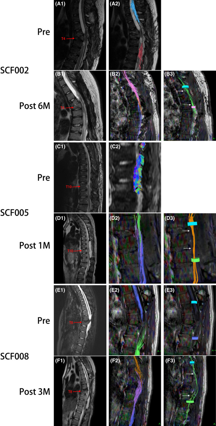
FIGURE 6.

Representative neuroimaging images of participants SCF002, SCF005, and SCF008 treated with vSCT. Preoperative T2‐weighted MRIs images show significant spinal cord injury (A1, C1, E1). DTI images show complete disruption of the spinal cord fibers (A2, C2, E2, E3). Postoperative T2‐weighted MRI images show the vascularized transplanted spinal cord in the original SCI area (B1, D1, F1). The three colored nerve fibers shown by DTI images represent nerve tracing and imaging of the distal and proximal spinal cord and the vascularized transplanted spinal cord (B2, D3, F3). DTI images show the reconnection of some nerve fibers to restore the nerve continuity of the spinal cord (B3, D3, F3). Pre, Preoperatively; Post 1 M, 1 month postoperatively; Post 2 M, 2 months postoperatively; 6 M: 6 months postoperatively
